The aim of this paper is to highlight the usefulness of this approach to the study of the vasculature. The stereological approach can provide access to the three-dimensional spatial framework of complex vascular beds. In this presentation, the application of this approach to a variety of vascular beds will be illustrated with examples including the nervous system (brain and spinal cord) and the reproductive system (endometrium). Stereology has been applied to a wide variety of problems in Biology in fields such as Neurobiology, Reproductive Biology and Cancer Cell Biology. In Biology, it provides a spatial framework upon which to lay physiological and molecular information. To date, the amount of data produced by this 67 method in fish fecundity laboratories is still, however, very limited due to the high work load 68 involved. The wide applicability of this approach is because it relies on basic facts of geometry and statistics. 65 stereology (Mayhew and Gundersen, 1996) because there is no longer any requirement to 66 assume particle shape, size and orientation. Holes can also be considered as structures.

In the stereological approach, the exact nature or function of the structure is not important.

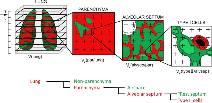
This approach allows inference of geometrical parameters such as volume, surface area, number, thickness and spacing. It may include any macro or microstructure in biology, materials sciences or indeed geology. The nature of the structure under study in itself does not matter. The study shows the various sources of non-reproducibility for deep learning results.The main objective of the stereological approach is to make estimations of parameters of geometrical structures using sampled information.
#Stereology mayhew pdf professional
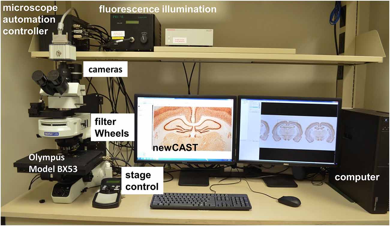
We also provide expert outsourcing for stereology analysis and professional tissue processing through our contract research organization (Stereology CRO). Moreover, the dissertation presents an experimental study of the effect of the non-reproducibility of deep learning models using two different deep learning libraries. SRC Biosciences (Stereology Resource Center, Inc.) is a well-established (25+ years) science and technology company that develops, commercializes and supports stereology systems (Stereologer®). This technique uses a previously trained deep learning model to create masks and query an unlabeled pool of data for masks to be verified by a human and increase the training examples for the deep learning model to improve performance. This contribution reviews the essential features of the stereological approach, identifies useful structural quantities, and provides examples of their application in various experiments of nature, particularly on normal gestation and the effects of pregnancies associated with high altitude, maternal diabetes mellitus, preeclampsia, and maternal smoking. Second, due to the limitations of the generalization ability of ASA and the growing amount of unlabeled data - due to the advancements and automation of microscopy image acquisition technology such as the Stereologer system, an algorithm to leverage unlabeled data using an active learning approach is proposed. First initial microscopic image labels (cell masks) were created using a handcrafted algorithm called the Adaptive Segmentation Algorithm (ASA), followed by human verification, instead of creating pixel-level cell masks manually. Here, we propose multiple approaches to generate and leverage unlabeled data for training deep learning models. Therefore obtaining labeled data remains a bottleneck for effective supervised leaning based models. However, labeling data is time-consuming, labor-intensive, and requires expert knowledge in many fields, such as neuroscience. These methods are based on supervised deep learning, which requires large labeled data sets for training in order to learn to count cells effectively. This dissertation provides deep learning methods to automate unbiased stereology cell counting in microscopy images of mice stained brain sections. Thus, the lack of high-throughput technology for automating unbiased stereology analyses remains a major obstacle to further progress in a wide range of neuroscience and cancer sub-disciplines.

#Stereology mayhew pdf manual
However, this human-based manual approach is time-consuming, labor-intensive, subject to human errors, recognition bias, fatigue, variable training, poor reproducibility, and inter-observer error. Unfortunately, in unbiased stereology current practices rely on a well-trained human to manually count hundreds of cells in microscopy images. The gold-standard for quantifying cells in tissue sections is the unbiased stereology approach. Cell quantification in histopathology images plays a significant role in understanding and diagnosing diseases such as cancer and Alzheimers.


 0 kommentar(er)
